Digital Mammography

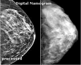
The standard mammogram that all women are encouraged to start having done yearly at the age of 40 is a film shot of the breast. The method has not changed significantly since mammography was introduced in the 1960s. The breast is compressed and an x-ray image is taken. In digital mammography, the x-ray source remains the same, but electronic detection provides vast improvements over film. An electronic image can be processed and color enhanced to provide the radiologist with tools that improve detection. Other very important advantages include the use of less radiation for a digital mammogram and better resolution on dense breasts, historically a problem for film.

Once a suspected area is located with full-field mammography, the suspect area can be resolved further using ultra-sound technology. Unfortunately, ultra-sound imaging has major disadvantages ranging from high false positive rates to the inability to detect some types of cancers. Spot digital mammography, an emerging field for the evaluation of lesions, micro-calcifications and suspected malignancies offers improved image resolution over the full-field image and ultrasound alternatives.
AEI was instrumental in the development of the first full field digital mammography with published clinical images and was subsequently involved in clinical trials. AEI was also involved in troubleshooting for the first spot digital mammography.
|